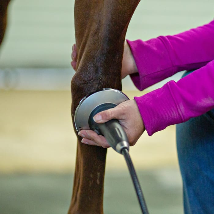

Extracorporeal Shockwave Therapy For Horses
Extracorporeal shockwave therapy (ESWT) has been documented to have regenerative and analgesic effects on bone and soft tissue.
Shock waves are high-energy acoustic waves that create micro trauma to increase blood supply, modulate inflammatory processes, stimulate regeneration, promote healing, decrease pain, increase the immune response in acute injuries and jump-start the immune system in chronic injuries.
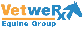
VetweRx Equine utilizes shockwave therapy for a variety of conditions.
Shockwave Applications
Below is a list of common applications for shockwave therapy:
- Desmitis
- Tendonitis
- Synovitis
- Bone cysts
- Caudal heel pain (navicular syndrome)
- Stable stress fracture
- Splint bone fracture
- Avulsion fracture
- Kissing spines (dorsal spinous process impingement)
- Lumbosacral pain
- Sacroiliac pain
- Muscle strains
- Periostitis
- Ringbone
- Sesamoiditis
- Splints (interosseous ligament tear)